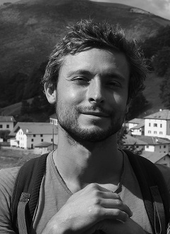
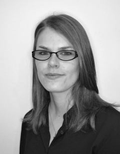
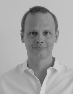
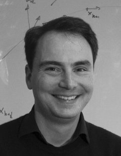
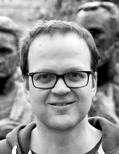
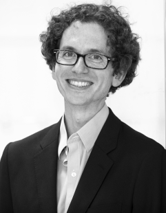
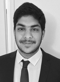
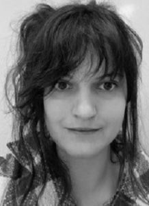
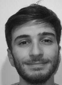
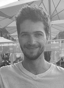
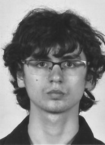
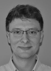
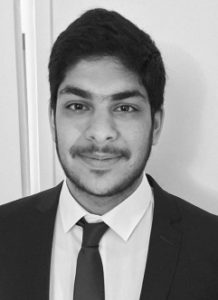
LAZARD Tristan
tristan.lazard [at] mines-paristech.fr
Short bio
Master 2 Mathematics and applications, UPMC
Thesis title
Deep learning for digital pathology: from full to no supervision.
Short abstract
Digital pathology involves studying digital versions of tissue slides to make diagnoses or derive prognostic markers from cells and tissue features. Deep learning can automate some diagnostic steps or help discover associations between phenotype and genotype. However, these slides are large, with full magnification versions weighing up to 16GB uncompressed, which therefore raises specific challenges. The goal of this PhD is to determine the best supervision methods to extract information from these slides.
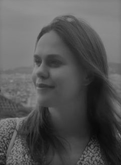
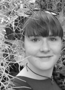
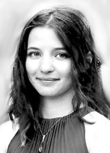
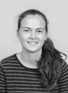
HEMFORTH Lisa
lisa.hemforth [at] icm-institute.org
Short bio
Master in Biomedical Engineering (BioImaging) from BME Paris (Arts et Métiers, Université de Paris, PSL, Télécom)
Thesis title
Deep learning for rating of atypical anatomical patterns on MRI data.
Short abstract
Incomplete hippocampal inversion (IHI) is an atypical anatomical pattern of the brain found in 15 to 20% of the general population which’s origins and link to different pathologies are still unknown. The aim of this project is to develop automatic rating methods based on anatomical criteria to apply them to big databases which would prove difficult to annotate manually. Using this data, we will perform genome wide association studies as well as heritability studies and study correlations to different pathologies.
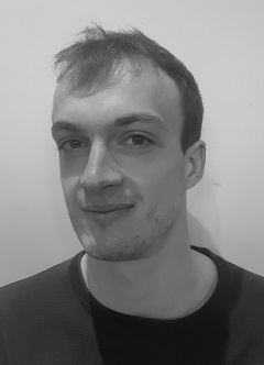
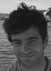
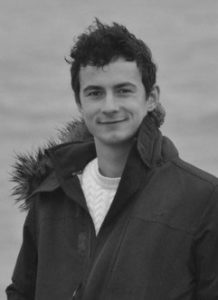
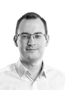
BONTE Thomas
thomas.bonte [at] mines-paristech.fr
SHORT BIO
Ingénieur Civil des Mines de Paris – Mines ParisTech
THESIS TITLE
Artificial intelligence for the analysis of label-free microscopy.
SHORT ABSTRACT
Deep learning algorithms outperform traditional methods for common image analysis tasks, with powerful architectures that have been developed recently. The goal of this PhD is to leverage these algorithms to provide tools for biologists in order to help them in their studies. Such deep learning methods will help them save time, avoid tedious annotation or simplify their experiments in their daily studies.
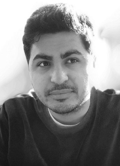
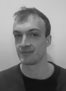
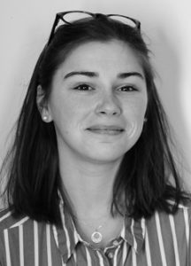
YAZDAN PANAH Arya
arya.yazdan-panah [at] icm-institute.org
Short Bio
Diplome d’ingénieur, Grenoble INP Phelma
Thesis title
Deep learning for the analysis of medical images in multiple sclerosis.
Short abstract
Multiple sclerosis (MS) is a chronic disease of the central nervous system with both an autoimmune and a neurodegenerative component. Conventional MRI techniques poorly predict disability worsening, leading to research exploring molecular imaging with PET and new biomarkers, such as choroid plexuses (CPs), which have been proposed as a neuroinflammatory biomarker along the entire disease course. This project aims to develop deep learning methods for automatic processing of MRI data to study CP segmentation and perfusion changes, as well as to explore the clinical course of patients based on the combination of different information.
Biomedical image analysis plays a crucial role in life sciences and healthcare, enabling researchers and clinicians to extract valuable insights from vast amounts of visual data. Automatic analysis of microscopy data enables quantitative studies of fundamental biological processes, such as gene expression or cell division, or to investigate the mechanism of action of drugs on cells and model organisms. Medical image analysis aims at providing tools for computer-aided diagnosis and prognosis, as well as the identification of biomarkers for treatment response.
The research we conduct inside PRAIRIE in this field involves the design and validation of new methodological approaches, with applications in the fields of neurological diseases and cancer. We work on a large variety of imaging modalities, such as light microscopy, histopathology, magnetic resonance imaging (MRI), positron emission tomography (PET). We aim at providing AI tools that can assist clinical decisions, widen access to cutting-edge care, allow progression towards precision medicine and a better understanding of diseases and biological mechanisms.
AI is particularly promising in biological and medical imaging but major challenges remain to be addressed. Training AI often requires large amounts of annotated data, which may be unrealistic in a biomedical context. Moreover, robustness is a critical issue. In particular, translation from highly-curated research or benchmark data to clinical practice is a major challenge. We therefore need methods that can work in a low-data regime, identify failures modes and account for changes in data distribution. Finally, we have more and more multimodal datasets, where different image modalities or image and omics data are available for the same patients. Integrating these data will therefore be one of the major challenges over the next few years.
In addition to these research activities, PRAIRIE chairs in biomedical imaging are strongly contributing to training in AI for audiences of various types, from engineers to MDs to future entrepreneurs.
Finally, in collaboration with other 3IA institutes, PRAIRIE also plays a major role in animating the community in the field of biomedical image analysis at a national level. Notably, we have created the first annual French conference on AI for biomedical imaging that gathers stakeholders across academia, clinical practice and industry.
VAILLANT Ghislain
Research Software Engineer
Université Paris Dauphine-PSL
ghislain.vaillant [at] icm-institute.org
Short bio
PhD, King’s College London
Research project
Clinica
Short abstract
Clinica is the software platform for clinical neuroimaging studies involving processing of multimodal data (imaging and phenotypic) for patients with cognitive diseases. My work consists in extending this platform to provide its functionalities as a service in the Cloud in order to serve a wider scientific audience.
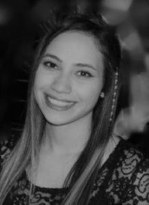
GODARD Charlotte
charlotte.godard [at] pasteur.fr
Short bio
- Engineer degree – Telecm Physique Strasbourg
- Master degree in Imaging, Robotics and Engineering for Healthcare – University of Strasbourg
Thesis title
Semi-automatic and amortized developments of transfer function for surgery planning in virtual reality.
Short abstract
Interpretation of medical images, such as MRI or CT-scan, can be challenging for a non-radiologist expert because of the various image quality and of the similarities between different structures of interest. However, surgeons need to understand these images to prepare surgeries and define corresponding anatomical landmarks. As universal segmentation is not possible due to the diversity of images between patients, we focused on the optimization of the visualization process applied only on the raw data. The AVATAR MEDICAL platform uses virtual reality for an intuitive visualization and manipulation of the images. Visualization parameters (color, transparency) are currently defined manually using an user-friendly transfer function desktop interface. The objective of the thesis is to automate the transfer function generation for a faster isolation of the structures of interest in the image, by combining a statistical approach and pre-trained models.
HASSANALY Ravi
rhassana96 [at] gmail.com
Short bio
Master degree (Diplôme d’ingénieur) at Ecole Centrale de Lyon
Thesis title
Deep generative models for detection of anomalies in the brain.
Short abstract
Neuroimaging offers an unmatched description of the brain’s structure and physiology, however identifying subtle pathological changes simply by looking at images of the brain can be a difficult task. The aim of this PhD project is to develop innovative computational imaging tools to model abnormalities, defined as deviations from normal variability, from multimodal brain imaging. To that purpose, deep generative models will be used to generate pseudo-healthy images from real patients’ images for different modalities. Comparing pseudo-healthy and real images will provide individual maps of abnormalities.
MASSON Jean-Baptiste
jean-baptiste.masson [at] pasteur.fr
Short bio
CSO of AVATAR MEDICAL, Principal investigator of the Decision and Bayesian computation laboratory, visiting scientist at Janelia research campus (2014-2019), visiting scientist at Institut Curie (2013), recipient of “Prix des innovateurs Ile de France” (2022) and the StartULM (2018), C’nano innovation (2017) and Young researcher price of the French Biophysics Society (2009).
Topics of interest
Bayesian inference, neuroscience, biological decision-making, statistical physics and data processing in virtual reality
Project in Prairie
Jean-Baptiste Masson will focus on Bayesian induction, structured inference, physical environment modeling and statistical physics to probe learning in insect brains using neuronal, connectome and behavior imaging. He will organize every two years symposium on links between neuroscience, AI and physics; and a graduate class on structured inference.
Quote
How are insects able to perform complex probabilistic tasks by leveraging only small small neural networks, whereas machine-learning tasks often require large-scale architectures and extensive training on massive datasets? Evolution is able to shape decision-making in small neural circuits while maintaining high performance. By joining physical modelling, supervised and unsupervised structured inferences, Bayesian induction and numerical simulations, we can probe how evolution programmed robust decision making in the “brain“ of insects. In turn, we can extract key neural circuits from these insects and test their performance in real-world tasks.
Team
BARRÉ Chloé
Postdoctoral researcher
BENICHOU Alexis
PhD Student
GODARD Charlotte
PhD Student
VERDIER Hippolyte
PhD Student
WALTER Thomas
Bioimage Informatics Computer Vision
thomas.walter [at] mines-paristech.fr
Short bio
Thomas Walter received his PhD from the Centre for Mathematical Morphology, Mines Paris-Tech. After 6 years of work at the EMBL Heidelberg, he joined the Centre for Computational Biology (CBIO, Mines ParisTech) in 2012. Since 2018, he is director of the CBIO and codirector of the department “Cancer and Genome: Bioinformatics, Biostatistics, Epidemiology of Complex Systems» (Institut Curie / Mines ParisTech / INSERM).
Topics of interest
Computer Vision, Bioimage Informatics, Histopathology, High Content Screening
Project in Prairie
Thomas Walter will work on problems in bioimage analysis. The main challenge he will address is to overcome massive annotations, which are often required for state-of-the-art computer vi-sion methods, but which seem unrealistic for many bioimaging projects. For this, he will investi-gate the experimental generation of ground truth data, weak supervision and image simulation. He will work on applications in fundamental cell biology, drug screening and histopathology.
Quote
Large-scale imaging approaches in biology and medicine are about to revolutionize basic life sciences and healthcare. Complementary to molecular approaches, they allow us to explore the spatial, morphological and multi-scale aspects of living systems. Artificial intelligence is the key technology today to transform this data deluge into knowledge. In this field, one of the major challenges for the next years is to overcome the need for massive annotation.
Team
BONTE Thomas
PhD student
Ingénieur Civil des Mines de Paris – Mines ParisTech
LAZARD Tristan
PhD student
Master 2 Mathematics and applications, UPMC
FOURNIER Laure
laure.fournier [at] parisdescartes.fr
Short bio
MD, PhD, Professor of Radiology, Hôpital Européen Georges Pompidou, Paris Cité University. Ecole Normale Supérieure (1991-1995), Research Fellow, UCSF, San Francisco, CA, USA (2001-2003). Responsible for the organisation of courses in Artificial Intelligence in Radiology for radiology residents, Member of the working group on Artificial Intelligence for the CERF – SFR, Member of the Scientific Committee of the DRIM France IA database. Grants over 650 k€ on radiomics and AI in medical imaging projects.
Topics of interest
Medical imaging, machine learning, radiomics, computer vision
Project in Prairie
Our project will focus on three approaches: 1) methodological developments on radiomics, i.e. high throughput extraction and selection of features from medical images using strategies including feature engineering, and deep learning and neural networks; 2) constitution of real-time prospective databases to obtain exploitable training and test data for the applications developed in Prairie; 3) integration of multimodality and multiparametric data stemming from multi-scale imaging going from microscopy to anatomical (radiology) and functional imaging.
Quote
Developments in computer vision need to translate into benefits for patients by transferring tools and applications developed for non-medical images to microscopic and macrsocopic medical imaging. The integration of this very diverse data to obtain a comprehensive view of a patient and his disease is a challenge which we must undertake in Prairie. The relative low numbers of patient data compared to the large number of features and parameters describing the patient and his disease, and the time-consuming annotation, remain important challenges and will require new tools which can train and learn on datasets with a weaker level of human supervision.
Team
DECOUX Antoine
PhD student
DURRLEMAN Stanley
Statistical learning, imaging, neurosciences
stanley.durrleman [at] inria.fr
Short bio
Stanley Durrleman is heading the joint Inria / ICM ARAMIS research team at the Brain Institute (ICM) in Paris. He is the founding director of the ICM centre for neuroinformatics. His research has earned him several international awards, including a European Research Council (ERC) award in 2015.
Topics of interest
Geometry and learning, neuroimaging, brain disorders, disease modeling, digital twins
Project in Prairie
We will develop novel statistical and computational approaches at the cross-roads of geometry and learning. These approaches built on generic principles will allow the exploitation of a large variety of structured and unstructured data such as clinical data, structural and functional imaging. These methods are well suited to deal with repeated data from the same patients over time (i.e. longitudinal data), so that they can be used to synthetize digital models of disease progression. The personalization of such models to new patient data will enable the implementation and evaluation of personalized therapeutic strategies.
Quote
Better understanding the brain and its disorders is probably the most fascinating scientific and medical challenge of this century. The repeated failures to find efficient treatments against most neurological diseases require to explore radically different approaches. At the core of one of the major European hospital and neuroscience research institute, we develop novel data-driven approaches to exploit large databases of neuroscience data including imaging, clinical, physiological and genomics data. We simulate the progression of neurodegenerative diseases. We design and evaluate decision support systems informed by personalized prediction of disease progression. Our research contributes therefore to the emergence of a precision medicine in neurology.
Team
DI FOLCO Cécile
Research engineer
POULET Pierre-Emmanuel
PhD student
SAUTY DE CHALON Benoit
PhD student
ORTHOLAND Juliette
PhD student
COLLIOT Olivier
Machine learning for medical imaging
olivier.colliot [at] upmc.fr
Short bio
Olivier Colliot is Research Director at CNRS and the founding head of the ARAMIS Lab, a joint team between CNRS, Inria, Inserm and Sorbonne University at the Brain and Spine Institute (ICM). Founded in 2012, ARAMIS gathers about 35 people dedicated to data science and AI for studying diseases of the brain. Prior to that, Olivier Colliot obtained a PhD in Computer Science from Telecom ParisTech in 2003, was a postdoctoral fellow at McGill University from 2003 to 2005 and joined the CNRS as a permanent researcher in 2006. He is the author of over 120 journal papers, an Associate Editor of Medical Image Analysis, one of the two leading journals of the field, and a Conference Chair at SPIE Medical Imaging.
Topics of interest
Machine learning, computer vision, medical imaging, multimodal medical data (imaging, genomics)
Project in Prairie
His research will be dedicated to interpretable machine learning for neuroradiology. The main research threads are: i) the design of approaches for more interpretable AI, ii) generic computer-aided diagnosis systems, iii) integration of multimodal data and iv) methodologies for reproducible research. Medical applications will be devoted to neurological diseases, in particular using large-scale clinical routine data.
Quote
Brain imaging is a domain in which AI hold major promises. However, current systems are not interpretable and too narrow. These are major barriers to their adoption by clinicians. I firmly believe that fundamental research advances are needed to make systems more interpretable and generic. I hope that these ultimately lead to better diagnosis and care of patients. I am really excited to conduct this project within PRAIRIE, which will open new fruitful collaborations with academic and industrial partners. I also believe we have a major role to play in training in AI the next generation of engineers but also clinicians, that our country needs.
Team
BRIANCEAU Camille
Engineer
Master degree (Diplôme d’ingénieur) at Institut d’Informatique d’Auvergne (ISIMA)
Master degree in imaging and technology for medicine (Université Clermont Auvergne)
FU Guanghui
PhD student
Master of Software Engineering; Beijing University of Technology
KMETZSCH Virgilio
PhD student
MSc in Data Science – Grenoble INP Ensimag & UGA
VAILLANT Ghislain
Research Software Engineer
PhD, King’s College London
YAZDAN PANAH Arya
PhD student
Diplome d’ingénieur, Grenoble INP Phelma
EL JURDI Rosana
Postdoctoral researcher
PhD from the University of Rouen Normandie
LOIZILLON Sophie
PhD student
Engineering Degree Bordeaux INP – Université de Bordeaux
HEMFORTH Lisa
PhD student
Master in Biomedical Engineering (BioImaging) from BME Paris (Arts et Métiers, Université de Paris, PSL, Télécom)
BURGOS Ninon
ninon.burgos [at] icm-institute.org
Short bio
Ninon Burgos is a CNRS researcher at the Paris Brain Institute, in the ARAMIS Lab. She completed her PhD at University College London, UK. In 2019 she received the ERCIM Cor Baayen Young Researcher Award, which is awarded each year to a promising young researcher in computer science or applied mathematics. Her research currently focuses on the development of computational imaging tools to improve the understanding and diagnosis of neurological diseases.
Topics of interest
Medical imaging, computer-aided diagnosis, machine learning
Project in Prairie
Ninon Burgos will focus on the individual analysis of medical images to improve differential diagnosis and strengthen personalised medicine. This project will involve developing advanced computational representations of multimodal imaging data and building flexible decision support systems. The framework will be applied to brain images to assist in the diagnosis of neurological diseases.
Quote
Neuroimaging offers an unmatched description of the brain, which explains
its crucial role in the understanding and diagnosis of neurological disorders. There is a critical need to develop new methods that can improve the characterisation of each patient’s pathology, and to build decision support systems more sensitive and easier to interpret.
Team
HASSANALY Ravi
PhD student
